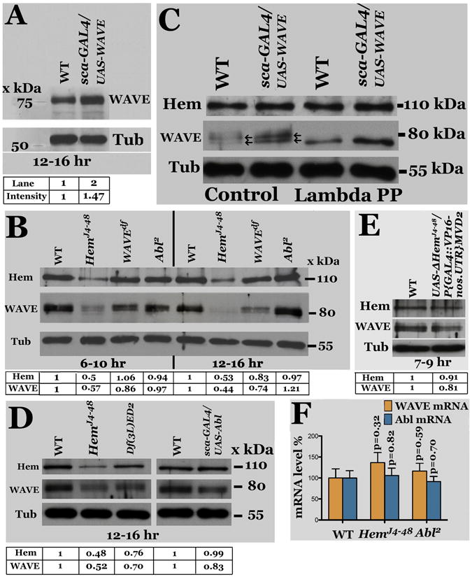
Figure 6. Regulation of WAVE by Hem.

Panel A: Western analysis for WAVE using anti- WAVE antibody on extracts from 12-16 hr old wild type and sca-GAL4/UAS-WAVE embryos. Panel B: Western analysis for WAVE, Hem and Tubulin (Tub, as loading control) from 6-10 and 12-16-hr old wild type (WT), HemJ4-48, WAVE-deficiency, and Abl2 embryos. Panel C: The WAVE protein is phosphorylated. Western analysis of untreated and Lambda phosphatase-treated embryo extracts for Hem and WAVE. Panel D: Western analysis of extracts from Hem and Hem-deficiency embryos for Hem and WAVE. Panel E: Western analysis for WAVE and Hem in wild type embryos expressing the truncated UAS-DHem transgene. Panel F: qPCR for WAVE and Abl mRNA from 12-16-hr old Hem and Abl mutant embryos. The error bars indicate standard error, with corresponding p values (student t-test). The intensity of the signals for WAVE and Hem, normalized against the loading control Tubulin, is given below each Western.
