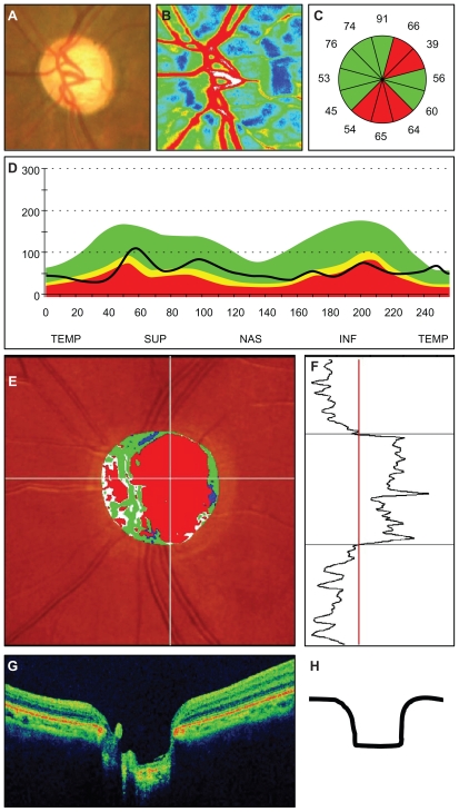
Figure 1.
Representative photographs of a patient with the generalized enlargement disc type of glaucoma. (A) Fundus photograph of the left optic disc. (B) Color map from LSFG-NAVI (Softcare, Ltd, Fukuoka, Japan). (C) Average thickness of the retinal nerve fiber layer thickness in the clock area. (D) Circumferential retinal nerve fiber layer thickness pattern in a patient with glaucoma. (E, F) Results of Heidelberg retina tomograph II. (G) Vertical section through the center optic nerve by three-dimensional optical coherence tomography.
Abbreviations: TEMP, temporal; SUP, superior; NAS, nasal; INF, inferior.

