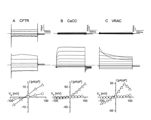
Figure 2.

At least three different Cl- channels exist in MAEC A. Current traces in a non-stimulated MAEC cell (upper traces) and in the same cell stimulated with the phosphorylation cocktail (lower traces) in response to voltage steps from +80 to -80 mV (decrement = 40 mV, VH = 0 mV). The bottom panel shows the corresponding I-V curves from the current amplitudes recorded at the end of each voltage step (open circles for the resting cell and open triangles for the stimulated cell). Note the voltage-independent kinetics of the current, the lack of rectification and the reversal potential close to ECl B. Current traces in an MAEC cell immediately after breaking into the cell and before it is loaded with Ca2+ (upper traces) and after equilibration with pipette Ca2+ (1 μM). Voltage steps form -100 to +100 mV (increment +20 mV), VH is -20 mV. Note the slow activation at positive potentials, and the inactivation at negative potentials, which are typical for CaCC currents. The corresponding I-V curves in the bottom panel illustrate the strong outward rectification of the CaCC current. C. Current traces from an MAEC cell before and during cell swelling induced by challenging the cell with a 25 % hypotonic solution. Same step protocol as in B. Note the inactivation of the current at positive potentials, which is a feature of volume-regulated anion channels (VRAC). The corresponding I-V curves at the bottom illustrate the weak outward rectification of the VRAC currents.
