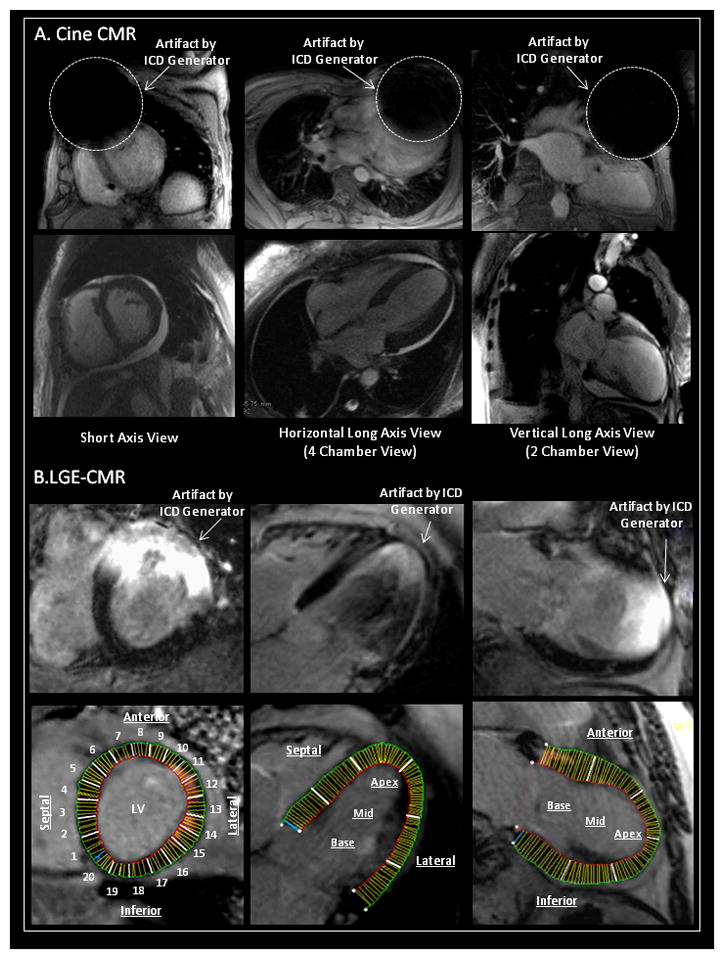
Figure 1. Methodology for Measurement of Artifacts on Cine CMR and LGE-CMR.

Artifact size on SSFP cine and late gadolinium enhanced cardiac magnetic resonance imaging (LGE-CMR) was measured in SA, HLA and VLA planes. (A) The artifact size on cine CMR due to the PM/ICD was measured. Percent sectors with any artifacts on cine CMR were also assessed in each plane. (B) The regional artifact effects on LGE-CMR due to the generator were quantitatively estimated in each plane (divided into 20 sectors in SA, 6 sectors in HLA, and 6 sectors in VLA planes, respectively).
SSFP=steady state free precession; SA=short axis; HLA=horizontal long axis; VLA=vertical long axis; PM/ICD=pacemaker and implantable cardioverter defibrillator.
