Figure 5.
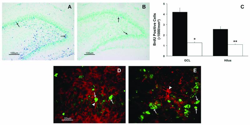
Bromodeoxyuridine (BrdU) immunohistochemistry of the dentate gyrus. Brdu positive cells in the granular cell layer (GCL) and hilus of the dentate gyrus of postnatal day 8 pups in the control (A) and morphine (B) groups are shown. Arrows point to BrdU positive cells. Morphine administration decreased the number of BrdU positive cells (C) in the granule cell layer and the hilus of the dentate gyrus on postnatal day 8. Values are mean ± SEM, n = 6 control (black), 8 morphine (white). * p<0.01 and ** p<0.05. Co-labeling of BrdU positive cells (green, thin arrow) with doublecortin (D, red, thick arrow) and glial fibrillary acidic protein (E, red, thick arrow) show that most BrdU cells co-label with doublecortin. Scale bar = 100 μm (A and B), and = 20 μm (D and E).

