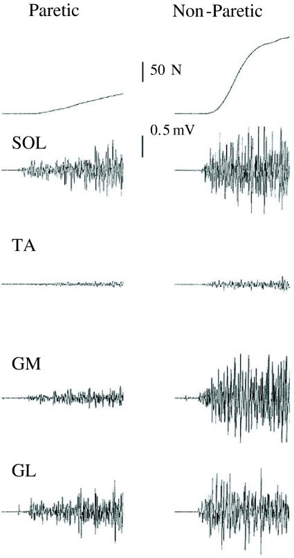
Fig. 4.
Recordings of plantar flexor maximum voluntary isometric contraction force and corresponding soleus (SOL), tibialis anterior (TA), gastrocnemius medialis (GM) and gastrocnemius lateralis (GL) electromyogram. The end of the sample traces are 500 ms after onset of contraction. The differences between the paretic and non-paretic limb can easily be observed

