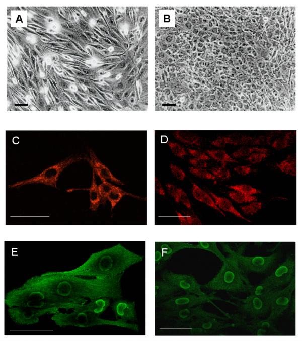
Figure 2.
GL15 phenotypes. Panels A and B represent, respectively, phase-contrast images of sub-confluent undifferentiated cells, grown in D-MEM containing 10% FCS, and differentiated cells incubated for 10 days in D-MEM supplemented with 2% HS. Panels C and D show, respectively, confocal GFAP localization in undifferentiated and differentiated GL15 cells immunostained with Cy3-conjugated anti-GFAP antibody (red fluorescence). The images (single focal plane at intermediate cell section) show no detectable difference in GFAP distribution between the two phenotypes. Panels E and F visualize, in undifferentiated and differentiated GL15 cells respectively, single focal plane at intermediate cell section images showing S100 expression in cells immunostained with OregonGreen-conjugated anti-S100B antibody (green fluorescence). The fluorescence signal indicates that S100B is localized in the perinuclear area. This finding is more evident in the differentiated phenotype. The same samples were also stained with TexasRed-conjugated anti-S100A, but no red fluorescent emission was detected. Bar = 50 μm.

