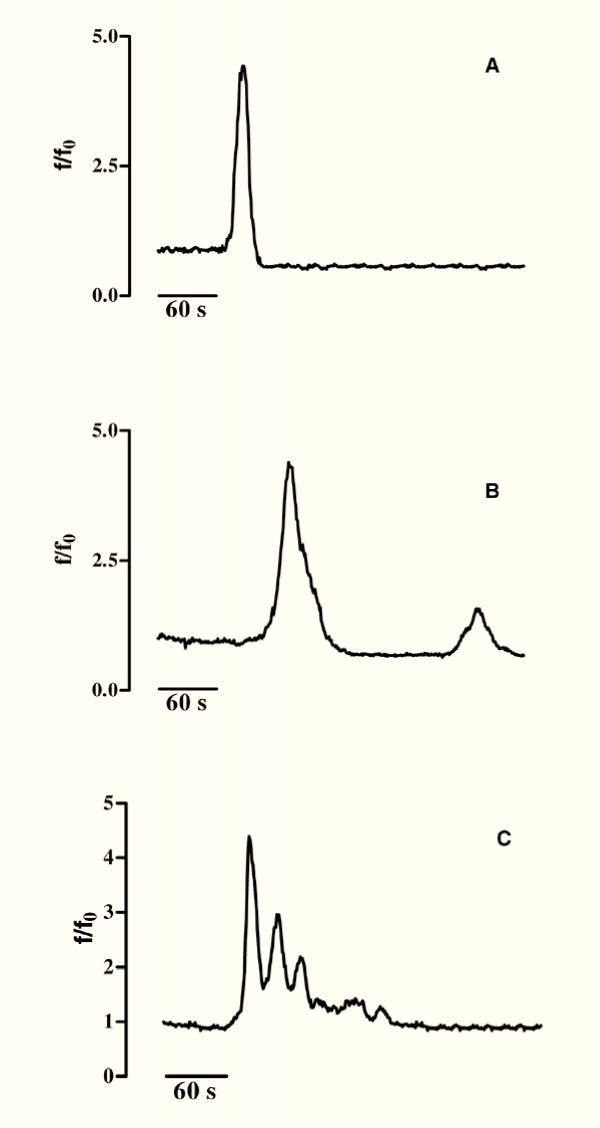
Figure 6.
Spontaneous [Ca2+]i variations in both undifferentiated and differentiated GL15 cells. Panels A, B and C show, respectively, a single [Ca2+]i spike, low frequency [Ca2+]i oscillations and high frequency [Ca2+]i waves. 50% (n>200) of the cell population (differentiated or not) seemed to prime spontaneous intracellular Ca2+ movements. Time (s=seconds) is indicated on the abscissa; the ordinate gives the normalized fluorescence value (f/f0).

