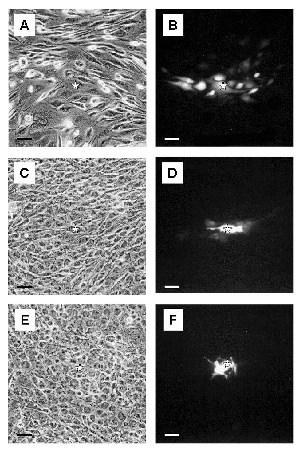
Figure 7.

Pattern of dye coupling in GL15 cell cultures Sub-confluent monolayers of undifferentiated GL15 cells (A and B) show the presence of dye-permeant junctional channels. When the monolayers reached confluence (C and D), the dye-spreading capacity of the cells was reduced. Almost no dye spreading is observed in differentiated confluent GL15 cultures (E and F). Fluorescence (B, D and F) and the corresponding phase contrast (A, C and E) photographs are taken on formaldehyde-fixed cells, 15 min after dye injection. The star symbol indicates the microinjected cell. Bar = 50 μm.
