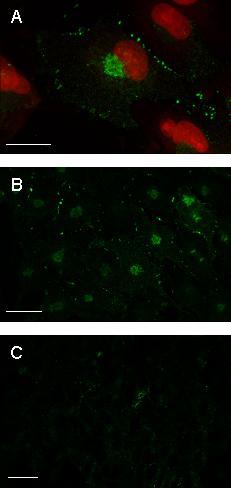
Figure 8.

Expression of cx43 in GL15 cells. Confocal microscopy image acquisitions of cells stained with anti-cx43 antibody revealed by OregonGreen-conjugated anti-IgG. In panels A sub-confluent undifferentiated GL15 cells are also stained with Propidium Iodide. The image shown in A is an X-Y projection of a tri-dimensional reconstruction of 12 sections. Confluent undifferentiated GL15 cells (B) appear to poorly express cx43 in respect to sub-confluent undifferentiated cells (A). This feature was also observed in the confluent differentiated phenotype (C). Panels B and C represent a single focal plane at intermediate cell section. Bar = 25 μm.
