
Figure 1.
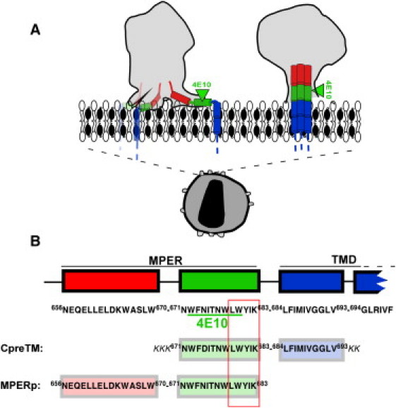
Models for gp41 MPER-TMD region organization at the HIV membrane and designation of the CpreTM and MPERp sequences used in this study. (A) Proposed models for the location of gp41 MPER-TMD at the Chol-enriched viral membrane. The cryoET density map contours of envelope spikes reported by Zhu et al. (5) and Zanetti et al. (12) are compared (left and right gray areas, respectively). Sequences depicted roughly parallel to the membrane plane (left) span amphipathic-at-interface and interfacial MPER subdomains (7). Cylinders inserted into the membrane represent the TMD anchors. (B) Sequences of the peptides used in this study. The diagram also shows positions and sequences for the potential cholesterol recognition/interaction amino acid consensus (outlined residue sequence) and 4E10 epitope (underlined residues). Sequence and numbering are according to the prototypic HXBc2 isolate.