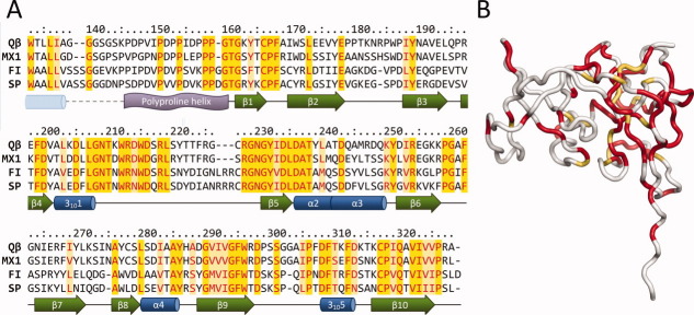
Figure 2.
Conserved regions of the read-through domains. (A) Sequence alignment of the read-through domains from different alloleviviruses. Conserved residues are colored red; of these, identical residues are shaded yellow and nonidentical light yellow. Assigned secondary structure elements are presented below the alignment. A dashed line represents the portion for which no experimental data are available; the α-helix from secondary structure prediction is drawn as a pale blue cylinder. (B) Mapping of the conserved regions on the three-dimensional structure of the read-through domain. Identical and nonidentical but conserved residues as of Figure 2(A) are colored red and yellow-orange, respectively.

