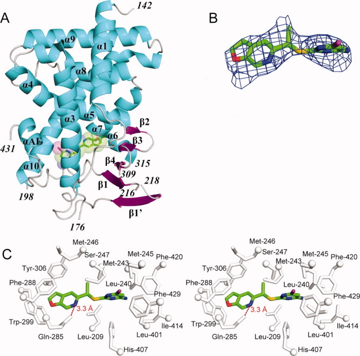
Figure 2.

Crystal structure of the human PXR-LBD in complex with PNU-142721. (A) The overall structure of PXR-LBD in complex with PNU-142721. The α-helices, β-strands and loops are rendered in cyan, magenta, and gray, respectively. PNU-142721 is colored by elements, with green, red, blue, yellow and magenta representing carbon, oxygen, nitrogen sulfur and chlorine atoms, respectively. Note the proximity of the PNU-142721 to αAF of the AF-2 surface of PXR. (B) The 2.8 Å simulated annealing omit map contoured at 2.5 σ is shown in blue mesh. PNU-142721 is shown and colored by elements. (C) Stereoview of the ligand-binding pocket of the PXR-PNU142721 complex. Residues that line the ligand-binding pocket of PXR are shown in gray and labeled. The hydrogen bond is represented by a red dash line. [Color figure can be viewed in the online issue, which is available at wileyonlinelibrary.com.]
