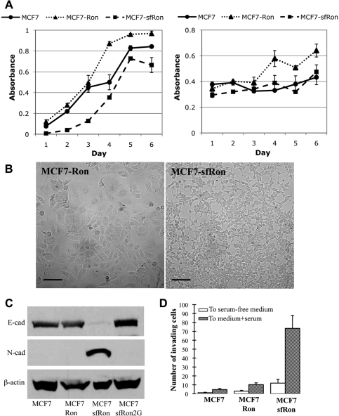
Figure 2.
sfRon confers EMT and invasive capability in breast cancer cells. (A) Left panel: proliferation of MCF7 and MCF7-sfRon cells based on mitochondrial dehydrogenase activity, read as absorbance over time. Right panel: survival of MCF7 and MCF7-sfRon cells in serum-free conditions based on mitochondrial dehydrogenase activity. (B) Phase-contrast micrograph illustrating altered morphology of MCF7 cells expressing sfRon (right panel) compared to those expressing full-length Ron (left panel). Photographs were taken at the same magnification; scale bars represent 100 µm. (C) Western blot with antibodies specific for E-cadherin, N-cadherin, and β-actin showed that sfRon decreased expression of E-cadherin and increased expression of N-cadherin. (D) sfRon induced invasion, as quantified by determining the number of cells that passed through Matrigel-coated Boyden chambers. No significant change was observed in MCF7-Ron cells. White bars: random invasion toward serum-free medium; gray bars: invasion toward medium containing 10% serum. Error bars reflect standard deviation from at least 3 experimental replicates.

