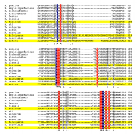
Figure 2.

Multiple amino acid sequence alignment of SB serine proteases. CLUSTALW was used to align amino acid sequences of SB serine proteases with experimentally determined and predicted 3D structures (highlighted in yellow). Only the regions showing the conserved catalytic residues Asp (D), His (H), and Ser (S) are shown. Amino acid residues with 100% conservation (*) between aligned sequences are either highlighted in blue (catalytic residues) or red (other). Other residues showing high (:) conservation (highlighted in gray) or medium (.) conservation are also indicated.
