
Figure 3.
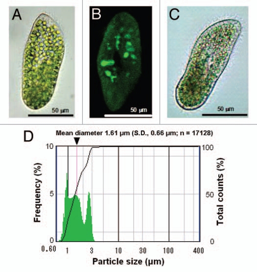
Continuous culturing of Synechocystis spp. PCC 6803 in the host cell. (A) Light microscopic images of wild-type P. bursaria. (B) Laser scanning confocal microscopic image of EmGFP-labeled E. coli-loaded cell of apo-symbiotic P. bursaria.2 (C) Light microscopic images of Synechocystis spp. PCC 6803-loaded cell of P. bursaria in the one-year-old symbiotic culture. (D) Statistical analysis of Synechocystis spp. PCC 6803 cells in 1 year-old symbiotic culture using a Symex flow particle image analyzer FPIA -2100.