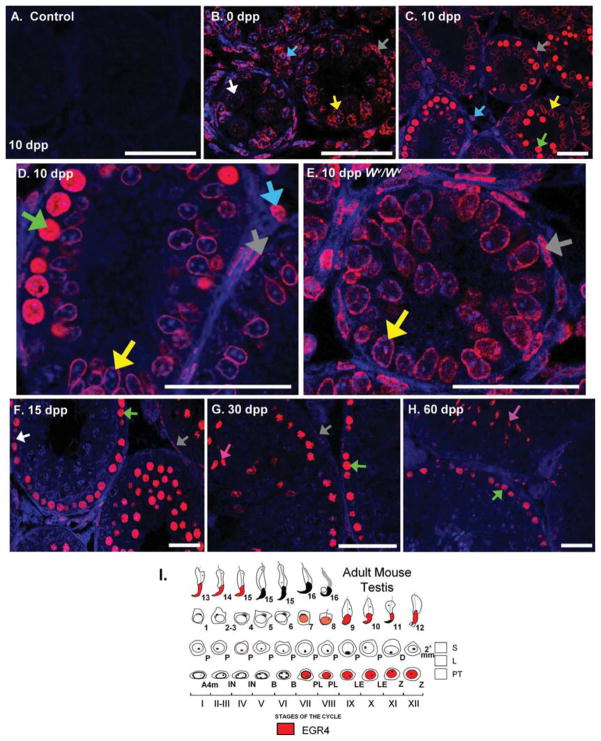
Fig. 3.
EGR4 protein localizes to germ and somatic cells in the developing testis. A–H: Immunofluorescence localizing EGR4 in sections of 0, 10, 20, 30, and 60 days post partum (dpp) wild-type mouse testes and to the 10 dpp Wv/Wv mouse testis. No primary antibody control shown in A. I: Mouse testis staging diagram detailing the cell expression and localization of EGR4 in the adult mouse testis. Scale bars = 20 μm. White arrows, spermatogonia; green, preleptotene/leptotene spermatocytes; pink, elongating spermatids; light blue, Leydig cells; yellow, Sertoli cells; gray, PTMs.

