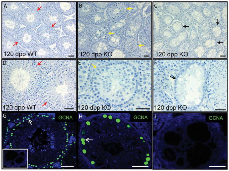
Fig. 5.

Egr4-null adult testis tubules regress with age. A–F: Hematoxylin stained sections of a 120 days post partum (dpp) wild-type (A,D) and Egr4-deficient (B,C,E,F) mouse testes. Red arrows denote normal tubules (A,D), yellow denotes partially regressed (B,E), and black denotes completely regressed tubules (C,F). G–I: Immunofluorescence localizing GCNA (green) to sections of Egr4 wild-type and null testes isolated from 1-year-old mice. A illustrates GCNA positive cells in tubules of a wild-type testis. Panel B depicts an Egr4-deficient testis tubule with multiple GCNA-positive cells at the basement membrane; however, C shows a tubule containing no GCNA-positive cells from the testis of the same animal. No primary antibody control on knockout tissue is shown as an insert in A.
