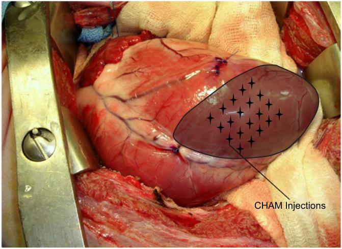
Figure 1.
Intra-operative photograph of an anteroapical MI as seen through a left thoracotomy. The infarct region is shaded and targeted injection sites of CHAM are noted by the stellate grid. Twenty injections were performed to a depth of 2 mm, placing a total of either 1.3 or 2.6 mL CHAM into the MI region.

