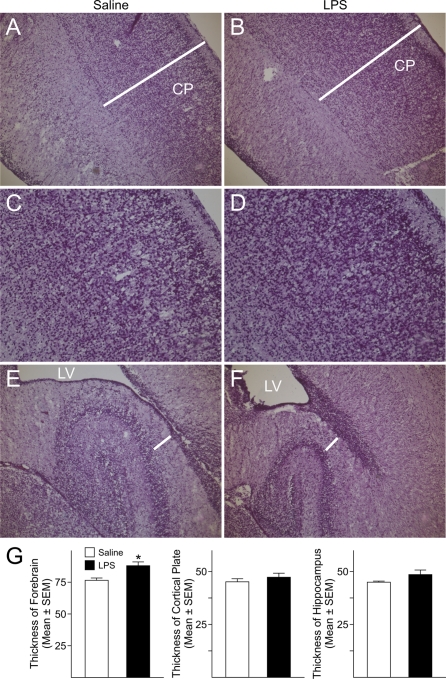
Figure 7. Changes in the offspring forebrain after prenatal foetal and maternal immune activation with LPS.
Dams received two consecutive injections of LPS (200 μg/kg, intraperitoneally) at GD15 and GD16 and the offspring were killed at postnatal day (P)1. (A, B) H&E staining revealed lack of significant differences in the thickness of the CP in P1 rats born to dams injected with saline or LPS; conversely, a significant enlargement of the cerebral cortex was found in P1 rats born to LPS-injected dams. (C, D) Higher magnification images showing increased cell density in the CP of P1 rats prenatally exposed to the effects of maternal injections of LPS. (E, F) No significant differences were found in the thickness of the hippocampus measured at the transition between the CA1 and CA2 areas. White bars indicate where the measurements were performed. LV, lateral ventricle. (G) Quantifications of the differences in the thickness of the cerebral cortex, CP and hippocampus. Histograms represent the means±S.E.M. for three P1 control (saline) and three P1 rats prenatally exposed to LPS. *P<0.05 versus saline-exposed P1 rats, Student's t test.

