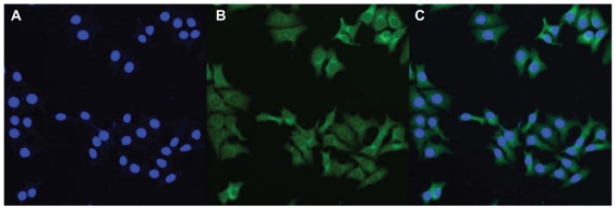
Figure 8.
Confocal laser scanning microscopy images of C6 cells after 2 hours of incubation with coumarin 6-loaded PCL-Tween 80 nanoparticles at 37.0°C. The cells were stained by DAPI (blue) and the coumarin 6-loaded nanoparticles are green. The cellular uptake was visualized by overlaying images obtained by green fluorescent protein filter and DAPI filter: left image from DAPI channel (A); center image from green fluorescent protein channel (B); right image from combined green fluorescent protein channel and DAPI channel (C).
Abbreviation: PCL, poly-ɛ-caprolactone.

