Figure 4.
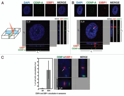
Centromeric regions are associated with persistent DNA damage foci in senescent hADSCs. (A) Immunofluorescent labeling of self-renewing hADSCs. Cells were seeded on coverslips and co-stained with anti-CENP-A (green) and anti-53BP1 (red) antibodies. DAPI staining is shown in blue. Confocal image of representative interphase nucleus is shown as separate channels and as a merged image. Four µm z-slice was analyzed by Imaris software and z1, z2 and z3 projections are shown. Self-renewing hADSCs show no focal damage associated sites. Centromeric areas are clearly visible. (B) Persistent DNA damage is associated with centromeres. Senescent hADSCs were seeded on coverslips and as in (A) and immunostaining was performed. An arrow depicts the co-localization of a centromeric region with persistent, senescence-associated γH2AX/53BP1 damage foci. Scale bar, 4 µm. (C) Quantification of CENP-A and 53BP1 co-localization in senescence. Senescent hADSCs were stained with antibodies against CENP-A (green) and 53BP1 (red) and DAPI (blue). Total of 200 cells were scored from three independent experiments. Error bars represent ± SAM. Example of higher magnification of the image is shown. Scale bar 1 µm. Images were analyzed by IMARIS software with optical sections representation as depicted on the left. Single 5 mm confocal section is shown. Image was analyzed by Imaris software and z1, z2 and z3 planes are shown. Cartoon demonstrates the orientations of z1, z2 and z3 planes within single z-section.

