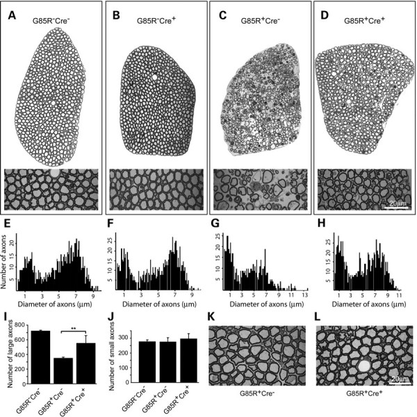
Figure 7.

Delayed motor axon degeneration in G85R SOD1/TrkB KO mice. (A–D) Toluidine blue staining of thin sections of L5 ventral root axons from 11-month-old animals showed substantial retention of large-diameter axons in G85R-SOD1+/−; VAChT-Cre+/− mice (D) compared with age- and sex-matched G85R-SOD1+/−; VAChT-Cre−/− mice (C). Elimination of TrkB alone (B) has no effects on axon morphology compared with wild-type control (A). (E–H) Nerve fiber diameter histograms from all the myelinated axons in L5 lumbar ventral roots showed preservation of two clear peaks and no consistent reduction in numbers of large-diameter fibers in the histogram from G85R-SOD1+/−; VAChT-Cre+/− mice (H) compared with G85R-SOD1+/−; VAChT-Cre−/− Mice (G). (I and J) Quantification of remaining axons from L5 lumbar ventral roots showed significant retention of large-diameter axons (>4 μm) in G85R-SOD1+/−; VAChT-Cre+/− mice (I, P < 0.01) compared with G85R-SOD1+/−; VAChT-Cre−/− mice. No change in the numbers of small-diameter axons (<4 μm) was seen (J). (K and L) Examination of 9-month-old animals showed normal appearance of axons in L5 lumbar ventral roots in G85R-SOD1+/−; VAChT-Cre−/− (K) and G85R-SOD1+/−; VAChT-Cre+/− (L) mice. Scale bars, 20 μm.
