Figure 1.

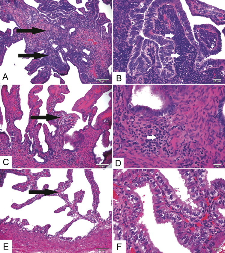
Representative fallopian tube C. trachomatis-associated histological findings at low (A, C, E) and high (B, D, E) magnification. In animal S5 (A and B), which met disease criteria for PID, the tubal propria (interstitium) was significantly and extensively thickened (long arrows) by numerous mononuclear cells, predominantly lymphocytes (short arrows). In animal S2 (C and D), which had 2 minor criteria consistent with infection but was not classified as PID, there was mild, localized expansion of the propria (long arrows) and scant lymphocytic inflammation in the propria and tubal wall (short arrow). In control animal S9 (E and F), the tubal propria was thin (long arrow) and lacked significant infiltrating inflammatory cells. Hematoxylin and eosin. Original magnifications panels A, C, E: 200×. Panels B, D, F: 600×.
