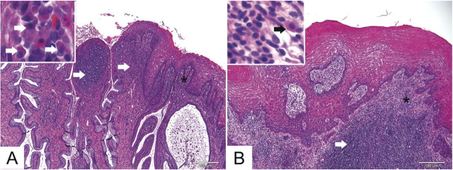
Figure 2.
Representative cervical and vaginal histological findings in C. trachomatis colonized (animal S7; A) and uninfected control (animal S10; B). Animal S7 (A), which had positive NAAT findings for 16 weeks, had robust lymphocytic and plasmacytic submucosal inflammation at the cervical squamocolumnar junction (long arrows). Inset shows plasma cells (short arrows) from area with asterisk. There was also extensive lymphocytic and plasmacytic submucosal inflammation in a control animal (S10; B) in the proximal vagina adjacent to the cervix (arrow). Inset shows plasma cell (short arrow) in area with asterisk. Hematoxylin and eosin. Original magnifications 100×.

