
Figure 1.
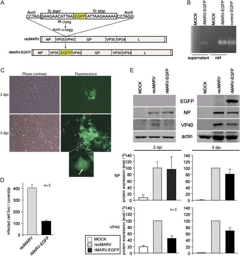
Characterization of recombinant MARV containing an ATU encoding EGFP. A, Scheme of rMARV-EGFP genome. The EGFP coding sequence is flanked by conserved MARV transcription start and stop signals. The EGFP ORF was inserted between the VP35 and VP40 gene via a newly created AvrII restriction site within the intergenic region (IR). The intergenic region spanning 5 nucleotides (CTATG) was altered to CCTAGG. B, Detection of recombinant genomes in supernatants and cell lysates of rMARV-EGFP-infected Vero E6 cells by RT-PCR. RT-PCR was conducted using primers binding in the EGFP ORF. Cellular RNA from Vero E6 cells transiently expressing EGFP was used as a positive control. C, Fluorescence microscopy of rMARV-EGFP-infected cells. Living cells were analyzed by phase contrast and fluorescence microscopy. Images were collected at 2 and 5 dpi. Bottom panel shows rMARV-EGFP-infected cell at higher magnification. Inclusions are indicated by an arrow. D, Comparison of progeny production of recombinant wild-type MARV (recMARV) and rMARV-EGFP. Vero E6 were seeded on glass coverslips and infected with recMARV or rMARV-EGFP. At 2 dpi, cells were subjected to immunofluorescence analysis using a MARV-specific antibody, and foci of infected cells were counted. The experiment was performed in triplicate and the bars represent mean values, including standard deviations. E, Quantitative Western blot analysis of virus protein and EGFP levels in Vero E6 cells infected with recombinant wild-type recMARV or rMARV-EGFP. Assays were performed in triplicate and standard deviations are shown.