
Figure 2.
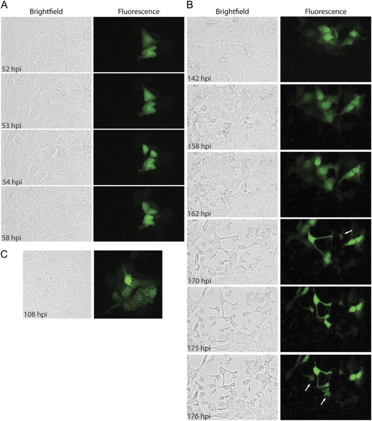
Time-lapse fluorescent microscopy of rMARV-EGFP spread. Vero E6 cells were infected with rMARV-EGFP at an MOI of 0.05, and EGFP fluorescence and phase contrast images were captured every hour for a period of 9 days. A, Cell division of infected cells. B and C, Cytopathic effects at late stages of infection. Blebbing cells are indicated by arrows. Time points postinfection when images were taken are indicated.