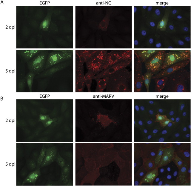
Figure 3.
Fluorescence microscopy analysis of EGFP and immunohistochemically labeled viral proteins in rMARV-EGFP-infected cells. Vero E6 cells were infected with rMARV-EGFP and subjected to immunofluorescence analysis at 2 and 5 dpi. Antibodies were directed against (A) intracellular viral proteins or (B) viral surface proteins. Antibody staining is indicated by red color; EGFP autofluorescence, green; and DAPI staining of the nuclei, blue.

