
Figure 4.
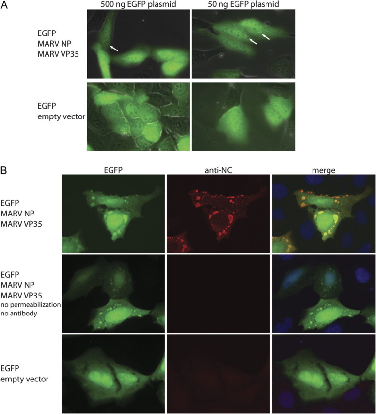
Accumulation of EGFP in MARV inclusions formed by NP and VP35. U2OS cells were transfected with an EGFP expression construct alone or along with plasmids encoding MARV NP and MARV VP35, as indicated. A, Live-cell imaging of transfected cells. EGFP autofluorescence is shown in green. Intracytoplasmic EGFP aggregates are indicated by arrows. B, Cells were stained using an antiserum directed against MARV nucleocapsid proteins (anti-NC; red) and DAPI (blue).