
FIG. 1.
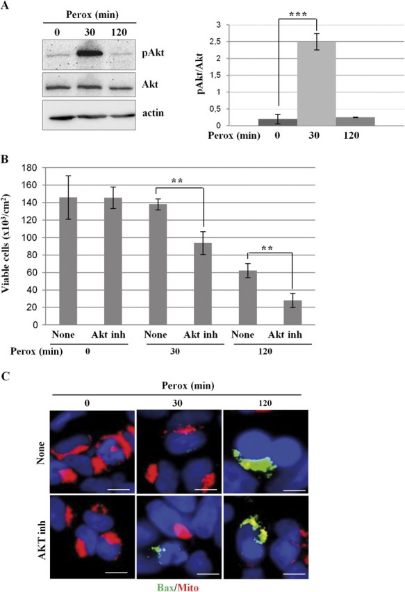
Inhibition of Akt sensitizes SH-SY5Y cells to H2O2 toxicity. (A) Western blotting of ser473-phosphoAkt and of total Akt in homogenates of SH-SY5Y cells exposed or not to 200μM H2O2 for the time indicated. One representative gel out of four independent experiments is shown. The filter was stripped and probed for actin as a marker of protein loading. The densitometry ratio of pAkt/total Akt is reported. (B) SH-SY5Y cells were preincubated or not with an Akt inhibitor and cultivated for up to 120 min in the absence or the presence of 200μM H2O2 (perox). At designated time-points, the adherent viable (trypan blue excluding) cells were counted. Cell density data obtained from three independent experiments in triple. (C) SH-SY5Y cells were preincubated or not with an Akt inhibitor and cultivated on sterile coverslips for 30 or 120 min in the absence or the presence of 200μM H2O2 (perox). At the end, the cells were stained with mitotracker and for immunofluorescence detection of active bax. Bax-positive/mitotracker-negative cells amounted to 15 ± 5% at 30 min and to 40 ± 15% (of the adherent cells) at 120 min in cultures exposed to H2O2 in the absence of the Akt inhibitor and to 42 ± 12% and to > 80% in the parallel cultures incubated in the presence of the Akt inhibitor. Data reproduced in three independent experiments. Bar = 10 μm.