
FIG. 4.
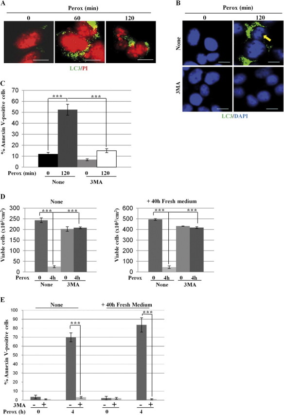
The Vps34 inhibitor 3-methyl adenine prevents H2O2 toxicity in SH-SY5Y cells. (A) SH-SY5Y cells plated on coverslips were exposed to 200μM H2O2 (perox) for 60 or 120 min and then fixed and processed for nuclei staining with propidium iodide (PI) and immunofluorescence detection of LC3-positive autophagosomes; 35 ± 5% of LC3-positive cells showed chromatin alteration typical of apoptotic cell death. (B) SH-SY5Y cells adherent on coverslips were preincubated or not with 3MA and exposed or not to 200μM H2O2 (perox) for 2 h. At the end, the cells were processed for nuclei staining with 4′,6-diamidino-2-phenylindole (DAPI) and immunofluorescence detection of LC3-positive autophagosomes. The arrow points to chromatin condensation and fragmentation in peroxide-treated cells. Bar = 10 μm. (C) Adherent cells preincubated or not with 3MA and exposed or not to 200μM H2O2 (perox) for 2 h. At the end, the cells were processed for cytofluorometry analysis of annexin V-FITC–labeled cells. Data (means ± SD) of four independent experiments. (D) Clonogenic assay to demonstrate the long-term protection by 3MA against peroxide stress. Two sets of cultures pretreated or not with 3MA were incubated for 4 h with or without H2O2 (perox), as indicated. At the end, the adherent viable cells of one set of cultures were counted. The cells of the other set of cultures were rinsed and cultivated for further 40 h in fresh medium and at the end the viable cells were counted. Cell density data (±SD) of two experiments in triple. (E) Cytofluorometric analysis of annexin V-FITC–positive cells of the cultures treated as above.