Figure 1.
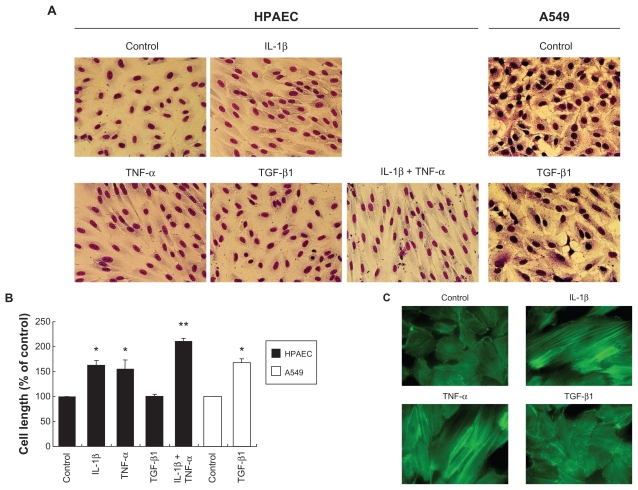
Effect of IL-1β, TNF-α and TGF-β1 on cell morphology, actin cytoskeletal arrangement. HPAECs and A549 alveolar epithelial cells were treated with 2 ng/mL of IL-1β, TNF-α, or TGF-β1 alone or in combination for 2 days. A) Cell morphology. Photographs were taken after Diff Quick staining (original magnification ×400). B) Cell size. Values are means ± SD of 3 separate experiments. C) Actin microfilament polymerization. F-actin was visualized by FITC-phalloidin (original magnification × 600).
Notes: *P < 0.05 compared with control; **P < 0.05 compared with cells treated with IL-1β.

