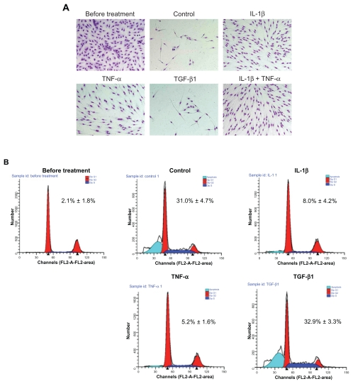
Figure 2.
Effect of cytokines on cell survival following withdrawal of growth factors. The subconfluent cells (before treatment, upper left panel) were cultured in serum and growth factor-free medium with or without 2 ng/mL of cytokines. A) Microscopic observation after 5 days of treatment. The cells remaining attached to the culture dish were fixed, stained, and photographed (original magnification ×200). B) Profiling of DNA contents after 2 days of treatment. Both floating and attached cells were collected and DNA content was profiled by FACS analysis. FACS tracings are from a single experiment. Insert: Percentage of apoptotic cells. Values are means ± SD of 3 separate experiments.
Note: *P < 0.01 compared with control.

