Figure 3.
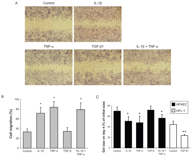
Effect of cytokines on wound closure and gel contraction. A) Microscopic observation of wound closure assay. Cells were treated with 2 ng/mL of cytokines for 2 days. After wounding, cells were incubated for 16 hours followed by microphotography as shown (original magnification ×40). B) Quantification of wound closure assay. Cell migration was quantified by counting migrated area at 0 and 16 hours after wounding. Data presented are means ± SD of 3 separate experiments. C) Gel contraction assay. HPAECs and human fetal lung fibroblasts (HFL-1) were cast into collagen gels (day 0) and treated with or without 2 ng/mL of cytokines for 4 days. Vertical axis: gel size expressed as percent of initial size (%). Data presented are means ± SD of 3 separate experiments.
Notes: *P < 0.05 compared with control; **P < 0.01 compared with control.

