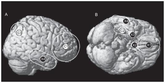
Figure 6.
Neural correlates of the C-system and the X-system displayed on a canonical brain rendering from side (A) and bottom (B) views. C-system regions displayed are the lateral prefrontal cortex (C1), hippocampus and medial temporal lobe (C2), and the posterior parietal cortex (C3). X-system regions displayed are the ventromedial prefrontal cortex (X1), nucleus accumbens of the basal ganglia (X2), amygdala (X3) and the lateral temporal cortex (X4). Please note that the hippocampus, nucleus accumbens and amygdala are displayed on the cortical surface for the sake of clarity. Copyright © 2004, American Psychological Association. Adapted from Lieberman MD, Jarcho JM, Satpute AB. Evidence-based and intuition-based self-knowledge: An fMRI study. J Pers Soc Psychol. 2004;87:421–435.

