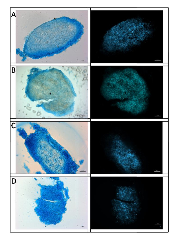
Figure 3.
Histology of MSC pellets 3 weeks post culture. Human mesenchymal stem cell (MSC) pellets were sectioned at 20 μm and stained with Alcian Blue (for GAG) and 4',6-diamidino-2-phenylindole (DAPI) (cell nuclei) after 21 days culture with four media groups; either B = Basal, C = Chondrogenic, NCA = notochordal NP cells in alginate; NCT = notochordal NP cells in tissue. (scale bar = 50 μm). GAG was observed for all media groups however NCT demonstrated the greater abundance of GAG throughout the whole pellet with fewer stained nuclei compared to other groups.

