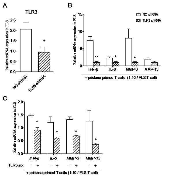
Figure 4.
Blockade of T-cell-activated FLSs via TLR3 signaling pathway intervention. Rat FLSs were transfected with TLR3-shRNA plasmid and a negative control. (A) TLR3 mRNA expression was detected 24 hours after transfection. TLR3-shRNA- and NC-shRNA plasmid-transfected FLSs were co-cultured with pristane-primed T cells, and (B) cytokine (IFN-β and IL-6) and MMP3 and MMP13 expression were detected at 24 hours. (C) Rat FLSs were preincubated with TLR3 antibody and isotype IgG, followed by pristane-primed T-cell co-culture for 24 hours and then expression of IFN-β, IL-6, MMP3 and MMP13 was detected. Data are presented as means ± SEM of four replicated determinations from three independent experiments. Levels of significance were calculated by using Student's t-test (*P < 0.05, **P < 0.01).

