Figure 4.
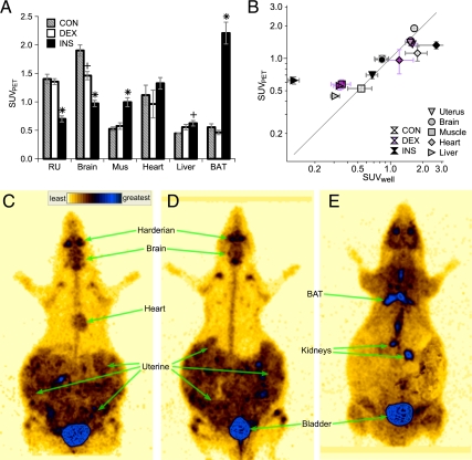
Final FDG disposition at the conclusion of PET imaging. A, SUVPET in indicated tissues in CON (gray stripe), DEX (white), and INS (black) groups. RU, Right uterine structures; Mus, skeletal muscle. +, ×, and *, P < 0.05 for difference vs. CON, DEX, and both, respectively, by ANOVA and Tukey’s HSD (n = 6–10 mothers per group). B, Correlation of SUVPET (y-axis) and SUVwell (x-axis). Tissues (right uterus, inverted triangle; brain, circle; muscle, square; heart, diamond; and liver, sideways triangle) and experimental groups (CON, white; DEX, purple; and INS, black symbols). C–E, Representative whole-body PET images for CON (C), DEX (D), and INS (E), highlighting regions of strong uptake. Heart is not distinctly visible and thus not labeled, in D and E; likewise for BAT in C and D. Regions directly exposed to FDG infusate, as defined on early frames, are masked.

