Fig. 2.
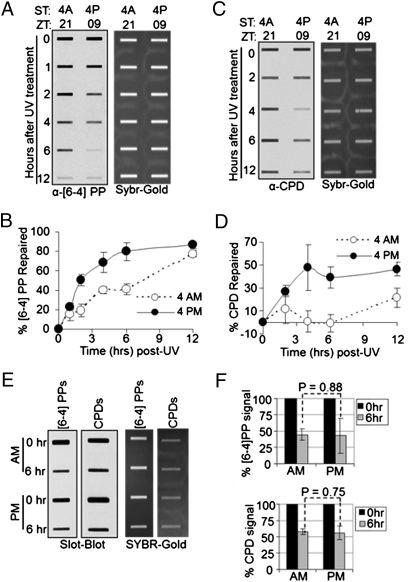
Circadian regulation of excision repair of UV-induced DNA damage in mouse epidermis. SKH-1 hairless mice kept under an LD 12:12 cycle were irradiated with 300 J/m2 of UVB at the indicated ZT times (either ZT 21 or ZT 09) and skin patches were collected 0–12 h after irradiation to measure UV photoproduct repair by slot blot. (A) Repair of (6-4) PP. A representative slot blot is shown. (Left) Immunoblot analysis of α-(6-4) PP signal. (Right) SYBR-GOLD staining of total DNA. (B) Quantitative analysis of (6-4) PP repair. Error bars indicate means ± SD (n = 2 mice at each time point). (C) Repair of CPDs analyzed by slot blot. (D) Quantitative analysis of CPD repair (n = 2 mice at each time point). (E) Slot blot analysis of (6-4) PP and CPD repair in Cry1−/−Cry2−/− mice. Skin was harvested at 0 and 6 h after irradiation and photoproduct levels were probed by immunoblotting. (F) Quantitative analysis of repair in the skin of Cry1−/−Cry2−/− mice irradiated at ZT 21 (4:00 AM) and ZT 09 (4:00 PM). The varied kinetics of repair in wild-type mice (A–D) presumably reflect the higher inherent rate of repair of the (6-4) PP and the cyclic expression of XPA. Error bars indicate means ± SD (n = 2 mice at each time point).

