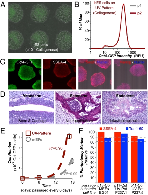
Fig. 4.
UV-patterned substrate supports standardized long-term culture. (A) Overlay of phase-contrast and fluorescent images of transgenic Oct4-GFP BG01 hESC cultures on “UV-Pattern” (as described in Fig. 2A) after 10 passages using collagenase dissociation. (B) Flow cytometry of cells after two consecutive passages on UV-Pattern. Max, maximum; p, passage; RFU, relative fluorescent unit. (C) Pluripotency marker immunostaining of cells on UV-Pattern. (D) Teratoma formation in immunodeficient mice by cells cultured on UV-Pattern. H&E staining was carried out on the teratoma, which contained tissues representing all three germ layers. (E) Number of undifferentiated Oct4-GFP–positive hESCs over three passages with accutase on UV-Pattern (red) and on standard mouse embryonic feeder (mEFs)-containing substrates (gray) when seeded with 24,000 cells per well of a six-well plate. Error bars indicate 95% confidence intervals (n = 3). High R2 coefficient of determination indicates good fit (R2 > 0.95) to exponential growth model (dashed lines). In A and E, the UV-Pattern was precoated with human serum. (F) Flow cytometry of cells for pluripotency markers SSEA-4 and Tra-1-60 after >10 consecutive passages on UV-Pattern for two different hiPSC lines, P237.1 and P237.5. Col, collagenase passaging. For the transgenic Oct4-GFP BG01 hESCs passaged on MEFs, only GFP-positive cells were analyzed for Tra-1-60 and SSEA-4 expression, allowing us to exclude MEFs from the analysis.

