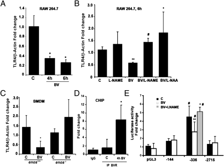
Fig. 4.
BV suppresses TLR4 expression dependent on eNOS. (A–D) Real-time PCR with primers for TLR4 in RAW 264.7 Mφ treated with (A) BV (50 μM) for 4 and 6 h. *P < 0.01, BV vs. control (C). (B) RAW 264.7 Mφ pretreated with L-NAME (100 μM) or L-NAA (10 nM) for 1 h before 6 h stimulation with 50 μM BV. **P < 0.004 vs. C; #P < 0.0004 vs. BV; *P < 0.04 vs. BV. (C) Enos+/+ and Enos−/− bone marrow-derived Mφ treated with BV for 6 h. *P < 0.03 vs. control. (D) ChIP assay in RAW 264.7 Mφ treated with BV (50 μM) for 4 h. *P < 0.02, BV vs. control (C). IgG was used as the negative control. Immunoprecipitation was performed with antibodies to BVR followed by real-time PCR with primers to the TLR4 promoter (−539 to −312 bp). Results represent mean ± SD of three independent experiments repeated in triplicate. (E) TLR4 promoter activity was measured in RAW cells that were transfected with the indicated luciferase reporter constructs comprising mutations of the TLR4 gene (27) for 24 h and cultured in the presence (white bars) or absence (black bars) of 50 μM BV for 4 h. L-NAME (100 μM) was applied 1 h before BV. Note that BV blocks luciferase expression in the absence of GATA-4 (−336 bp) but not Ap-1 (−144 bp). Results are presented as fold change over empty vector control and represent mean ± SD of three independent experiments repeated in triplicate. #P < 0.001 vs. pGL3; **P < 0.001 BV (−336 bp) vs. DMSO (−336 bp); *P < 0.0008 BV vs. BV + L-NAME.

