Fig. 5.
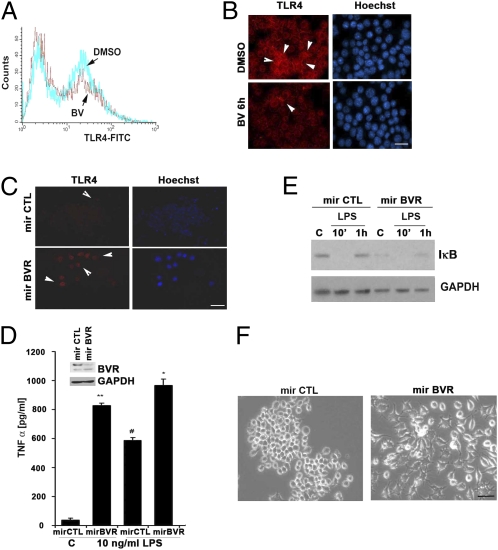
Lack of BVR leads to a proinflammatory phenotype in Mφ. (A) Flow cytometry of TLR4 FITC levels on the surface of RAW 264.7 Mφ treated with DMSO or BV (50 μM) for 6 h. Data are representative of at least three independent experiments. (B and C) Representative immunofluorescence staining of TLR4 in RAW 264.7 Mφ treated with BV or transfected with mirBVR or mirCTL controls to inhibit expression. Red, TLR4; blue, Hoechst. Images are representative of 10 fields from two independent experiments. Note that BV reduced, whereas blockade of BVR induced TLR4-positive staining. (D) ELISA for TNF-α levels in media collected after 24 h from RAW 264.7 Mφ ± mirBVR knockdown. Data represent mean ± SD of three independent experiments repeated in triplicate. *P < 0.0001 mirBVR vs. mirCTL; #P < 0.001 mirBVR + LPS vs. mirCTL + LPS; **P < 0.001 mirBVR vs. mirCTL. (Inset) Immunoblotting with antibody against BVR in cells stably transduced with mirBVR. (E) Immunoblot total IκB in mirBVR-expressing RAW 264.7 Mφ treated ± LPS (100 ng/mL) for 10 min and 1 h. Blot is representative of three independent experiments. (F) Morphologic changes in mirBVR-expressing RAW 264.7 Mφ vs. LMP-transfected control RAW 264.7 Mφ 24 h after seeding. Note that lack of BVR results in the activation of Mφ assessed morphologically. Images are representative of 10 fields of view in triplicate.

