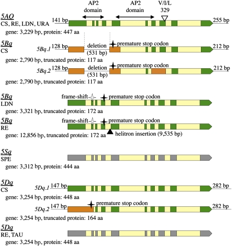
Fig. 3.
An overview of the intron/exon structure of the Q/q homoeoalleles determined by RT-PCR cloning and sequencing. Exons conforming to the splicing pattern found for the 5AQ gene are shown in green (deduced from cDNA sequences) and gray (predicted), alternative exons are shown in orange, and introns are shown in yellow. 5′- and 3′-UTR lengths (L) by RACE are shown for CS mRNAs. Gene sizes shown are from the translation start to the stop codon. Exons encoding the AP2 domains and the position of the codon for amino acid residue 329 in 5AQ (I), and the corresponding codons in 5Aq (V) and 5Dq (L) are also shown.

