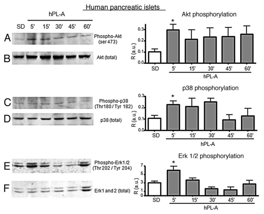
Figure 4.
Western blot analysis of proteins involved in the signal transduction pathway induced by hPL-A in human pancreatic islets. 90% clinical purity-grade human islets were serum deprived (SD) and incubated at different times (5, 15, 30, 45 and 60 min) with 500 ng/ml of hPL-A. Islets were then lysed and 50 µg of protein lysates were separated by 10% SDS-PAGE. The blotted proteins were incubated with the specific antibody anti-phospho-Akt, anti-phospho-p38 and anti-phospho-Erk1/2 (lane A, C and E, respectively). The protein amount was normalized utilizing the total antibody (lane B, D and F, respectively). Beside each couple of blots (A/B, C/D and E/F) we showed the relative densitometric analysis (R, Phosphorylated protein content/Total protein content; a.u., arbitrary units); *p < 0.05 vs. SD (n = 3). One representative blot of three different experiments performed with islets from three different donors is visualized.

