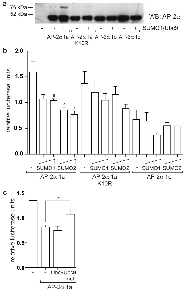
Figure 5.
AP-2α isoform 1a can be sumoylated leading to decreased transactivation activity. (a) HepG2 cells were transfected with 0.3 μg/well of the different pcDNA3-AP-2α constructs, without and with 0.1 μg/well pSG5 Ubc9 and 0.6 μg/well pSG5 SUMO1 in a six-well format. 48 h after transfection, lysates were harvested in RIPA buffer containing IAA and NEM, and analysed by western blot with a pan AP-2α antibody (3B5). One experiment representative of three is shown. A second, weaker sumoylation site (IKKG) was predicted (using "SUMOplot") within the C-terminal half of the protein which could explain the low levels of sumoylation observed for AP-2α K10R and the other isoforms using longer exposures. (b) HepG2 cells were transfected with 0.05 μg/well of the different pcDNA3-AP-2α constructs, 0.25 μg/well 3xAP2-Bluc, 0.25 μg/well phRG-renilla, 0.25 μg/well pcDNA3-CITED2, 0.75 μg/well pCI-p300, and 0.25 or 0.5 μg/well pSG-SUMO1/2 as indicated. Relative firefly luciferase activity normalised to renilla luciferase activity is shown. The average and standard error of three experiments is reported. (c) HepG2 cells were transfected with 0.2 μg/well pGL4.74 (Renilla), 0.3 μg/well cyclin D3 reporter, 0.15 μg/well pcDNA3-AP-2α isoform 1a, 0.3 μg/well pSG-Ubc9 and 0.15 μg/well pSG-SUMO1. Average and standard error from three independent experiments is represented.

