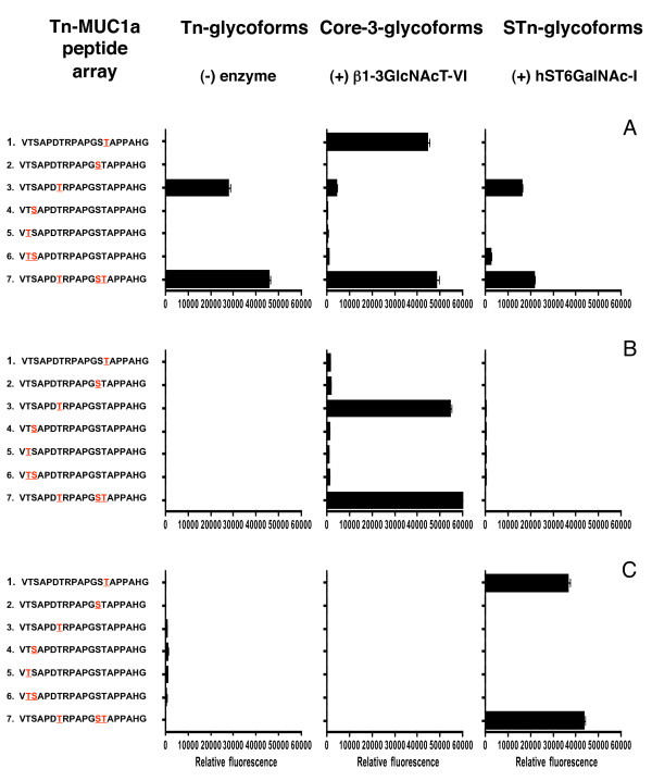
Figure 2.
Epitope mapping of autoantibodies in sera from breast cancer patients. Arrays were produced by on-slide glycosylation of the MUC1a glycopeptides carrying Tn at the sites indicated in red using core3 β3GlcNAc-T6 (middle panel) and ST6GalNAc-I (right panel). The arrays were stained with diluted sera (1:20) and Cy3 labelled anti-human-IgG. A, B, C show data obtained with sera from three individual patients.

