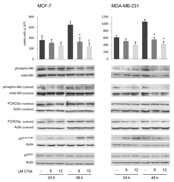
Figure 6.
Cell viability and immunoblot analyses in CTet-treated MCF-7 and MDA-MB-231 breast cancer cell lines. Cells were treated for 24 and 48 hours with 6.0 and 12.0 μM CTet or vehicle only (γ-cyclodextrin aqueous solution). After treatments, cell viability was evaluated by Trypan blue dye exclusion assay and cell extracts were processed as described in the Materials and methods ('Immunoblot analysis'). Akt activity was analyzed by using a phospho-sensitive Akt antibody in total cell extracts and in cytosolic cellular fractions. FOXO3a localization was evaluated by separation of nuclear and cytosolic proteins, and p21 and p27 overexpression was evaluated in total cell extracts. FOXO3a, p27, and p21 were normalized to actin, and phospho-Akt was normalized to total-Akt. Cell counts are presented as the mean ± standard error of the mean of three separate experiments. Asterisks indicate statistically significant values with respect to untreated cells (one-way analysis of variance followed by Tukey post hoc test; P < 0.01). Blots are representative of at least two separate experiments. CTet, indole-3-carbinol cyclic tetrameric derivative.

