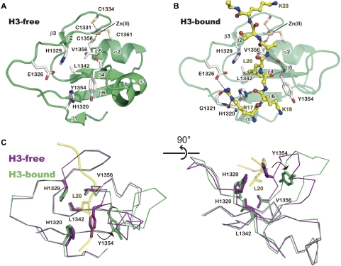
Figure 4.
Conformational change of the zinc-binding domain to accommodate the H3 peptide. (A) Structure of the zinc-binding domain in the H3-free structure. Secondary elements are labeled as in Figure 1. Purple dashed lines represent bonds coordinating Zn(II). (B) Structure of the zinc-binding domain bound with the H3 peptide. (C) Orthogonal views of the H3-free (purple) and H3-bound (green) structures of the zinc-binding domain. The H3 main chain (ribbon) and the L20 side chain (ball and stick) in the H3-bound structure are shown in yellow.

