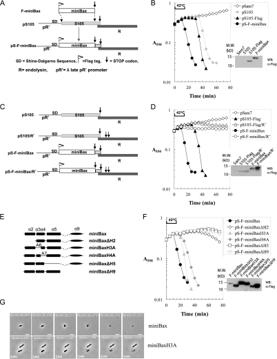
Figure 2.
MiniBax functionally replaces bacteriophage holin (S105), causing endolysin- dependent lysis of Δ(SR) cells. (A) Diagram for the construction of the pS105-based isogenic plasmid replacing S105 with F-miniBax. The DNA segment encoding the first 92 amino acids of S105 in the S105/R lysis cassette within the plasmid pS105 was replaced by the reading frame of F-miniBax. (B) Lysis of Δ(SR) cells by the miniBax/R cassette. The plasmid pSam7 is identical to pS105 except for an Amber mutation in the S105 gene that abolished S105 expression. Upon reaching A550 ∼0.3, the Δ(SR) cells carrying the indicated plasmids were induced by thermal induction for 15 min at 42°C, followed by aeration at 37°C. A550 was measured for each culture at 5-min intervals. The cultures were harvested as described in the Materials and Methods and subjected to SDS-PAGE and Western blot with an α-Flag antibody. (C) Diagram of the lysis cassettes of S105 and F-miniBax with or without functional endolysin (R). (D) Lysis phenotypes of the lysis cassettes listed in C in Δ(SR) cells. Expression of the indicated proteins was detected by Western blot with an α-Flag antibody. (E) Diagram of truncation mutants of miniBax. These miniBax mutants were cloned into the pS105 plasmid by the same strategy as shown in A. (F) Lysis induced by miniBax and its truncation mutants in Δ(SR) cells was measured the same way as in B. The expression of these mutants was detected by Western blot with an α-Flag antibody. (G) Δ(SR) cells carrying either pS-F-miniBax or pS-F-miniBaxH3A were monitored by time-lapse microscopy. Still images of the cells undergoing lysis at different time points are shown. The real-time videos are provided in Supplemental Movies S1 and S2.

