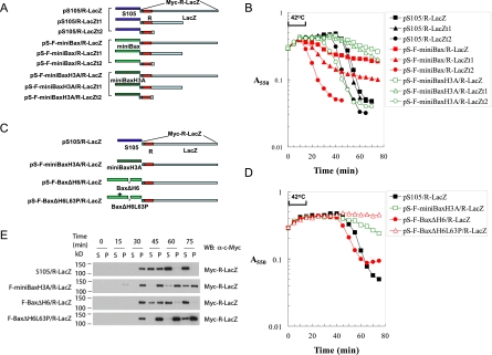
Figure 6.
Sizing the membrane holes induced by active Bax mutants through the R-LacZ fusion proteins. (A) Diagram of the various lysis cassettes with LacZ or its truncation mutants fused to the reading frame of R. The R-LacZ fusion protein has a Myc tag on the N terminus. R-LacZt1 is the fusion between R and the first 497 amino acids of LacZ. R-LacZt2 is the fusion between R and the first 8 amino acids of LacZ. (B) Lysis curves of the different lysis cassettes listed in A in Δ(SR) cells. (C) Diagram of the lysis cassettes expressing Bax mutants in conjunction with R-LacZ. (D) Lysis curves of the indicated Bax mutants with R-LacZ cassettes in Δ(SR) cells. (E) Δ(SR) cells carrying the indicated plasmids were thermally induced. At the indicated time points, 1-mL samples of the culture were collected and pelleted by centrifugation at 22,000g at 4°C. The supernatant (S) and the pellet (P), which was resuspended in 1 mL of EBC buffer, were loaded onto SDS-PAGE followed by Western blot analysis with an α-Myc antibody.

